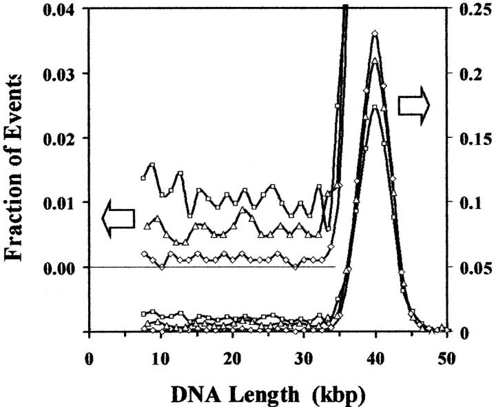
FIGURE 8.
Histograms for DNA from bacteriophage T7 that has not been irradiated (⋄), irradiated with a dose of 1 Gy of γ-rays from a 137Cs source (▵) and irradiated and then treated with D. radiodurans Fpg protein that makes single-strand breaks at certain oxidized purines (□). To facilitate comparisons, the histograms are plotted as a function of molecular length and have been normalized by dividing by the total number of peaks recorded for each histogram so that they have the same area and the vertical axis becomes the fraction of events recorded per binning interval for each sample. The right scale represents the events for the main plot, while the left scale is expanded by a factor of five and offset by 20% to facilitate comparisons of the low number of events recorded for smaller molecules. The data show that radiation alone decreases the fraction of the events corresponding to intact T7 DNA molecules and increases the events corresponding to fragments of T7 DNA. Subsequent treatment with Fpg protein produces a further drop in the fraction of intact molecules and an increase in the fragments. In the case of the untreated DNA and the irradiated-only sample, the distribution of samples is, within experimental uncertainty, independent of fragment length, whereas for enzyme-treated sample, the smaller fragments may occur at a slightly higher frequency, reflecting an observable rate of multiple breaks in some of the intact molecules.

