FIGURE 2.
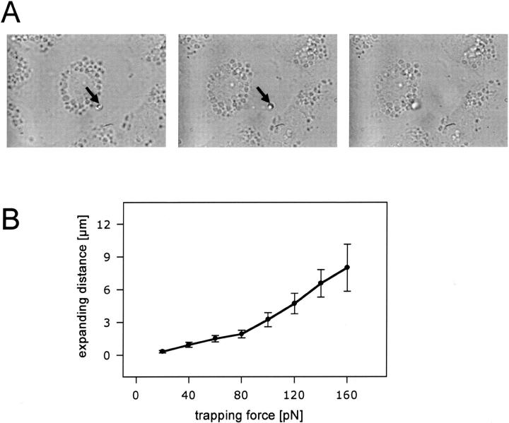
(A) Expansion of exocytosed surfactant. Transmission microscopy images demonstrating extension of a single FM 1-43-labeled fused LB. The fused LB was trapped in the laser beam (arrows indicate the position of the laser trap). (Left), Trapped LB before movement of the translational stage (i.e., before cell movement). (Middle), Trapped LB after movement of the stage. Note the extension of surfactant. (Right), Untrapped LB (laser power turned off) following extension. Note the “recoiling” of surfactant. (B) Correlation between trapping force and expansion length of fused LBs. Dots indicate mean ± SE of the mean (n = 34).

