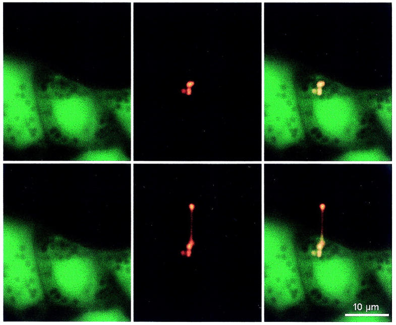
FIGURE 3.

Differential staining of the cytoplasm (Calcein, left images) and of surfactant from fused vesicles (FM 1-43, middle images), in a cell before stretch (upper images), and during stretch (lower images) of a fused LB. The right images represent the overlay of the Calcein and FM 1-43 stainings. Before the experiment, cells had been preincubated for 30 min in 1 μM Calcein. Note that three neighboring vesicles stain with FM 1-43, suggesting compound exocytosis at this site. Only one of these three fused vesicles could be pulled by the laser trap.
