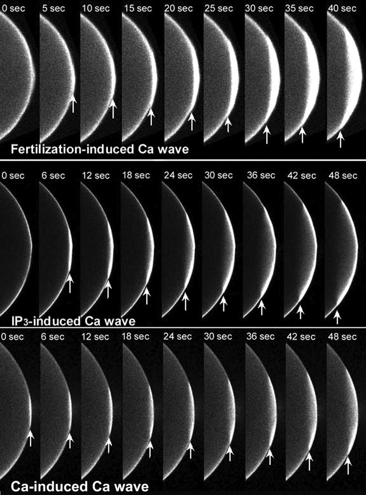
FIGURE 1.

Time sequence of confocal images of Ca2+ wave initiation using a 40-μm thick optical section. Albino Xenopus eggs were injected with calcium green-dextran and activated by sperm (top), IP3 microinjection (middle) or Ca2+ leak (bottom). Arrows mark the leading edge of the Ca2+ wave. Both fertilization and the microinjection of IP3 produced a Ca2+ wave that traveled faster during the first 10–15 s than waves triggered by a Ca2+ leak.
