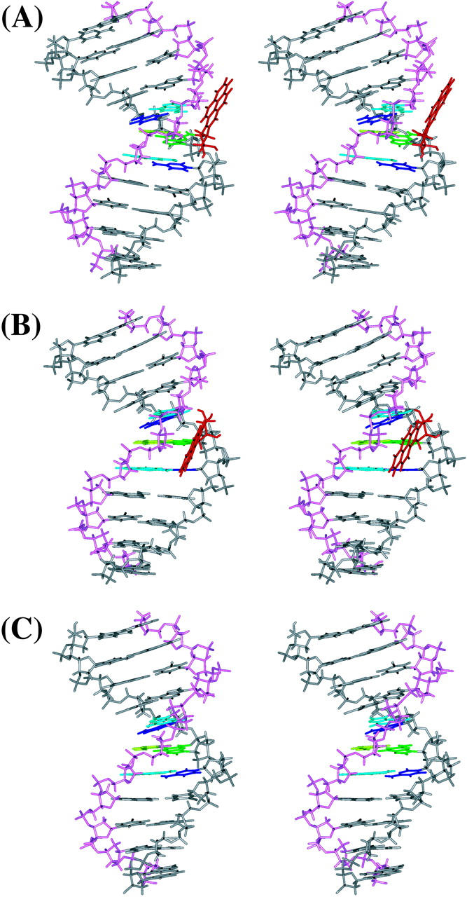
FIGURE 3.

Stereo views of the MD average structures for (A) 10S (+)-trans-anti-dG adduct, (B) 10R (−)-trans-anti-dG adduct, and (C) unmodified control duplex over 1.5–3 ns. BP is red, G*6 (G6 in the unmodified control) is green, T17 is light green, C5 and C7 are blue, A16 and A18 are light blue, and other residues are gray. The backbone, sugar atoms from C1 to C11 are gray, and those from G12 to G22 are light magenta. All structures are aligned to have 5′-OH at the top left. All stereo images are constructed for viewing with a stereo viewer.
