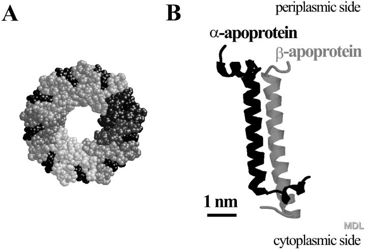
FIGURE 1.
(A) Topology model (top view, periplasmic side) of the LH2 of Rps. acidophila strain 10050. It consists of a concentric arrangement of two hollow cylinders built from nine α- and nine β-apoproteins. The outer surface of the cylinder is formed by the β-subunits, and its inner surface by the α-subunits. Each αβ-apoprotein pair binds four pigment molecules, three bacteriochlorophylls, and two carotenoid molecules. The spheres in various shades of gray represent the 18 apoproteins and the black spheres represent the pigment molecules. (B) Topology model of an αβ-apoprotein pair without the associated pigment molecules. Model A has been scaled down five times with respect to Model B.

