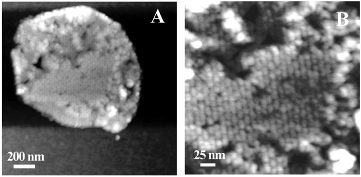
FIGURE 3.
(A) TMAFM topograph of reconstituted light-harvesting 2 complex in a lipid bilayer imaged under ambient conditions. The gray scale represents a height range of 15 nm. (B) The higher magnification image shows periodic structures, with an ellipsoidal-like shape. The gray scale represents a height range of 5 nm.

