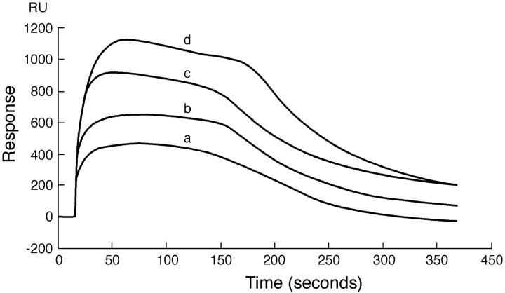
FIGURE 1.
Sensogram depicting the binding of PDC-109 to pure DMPC monolayer at 20°C. PDC-109 in varying concentrations was passed over a monolayer of DMPC: (a) 0.75 μM, (b) 1.0 μM, (c) 1.5 μM, and (d) 3.0 μM. The sensogram obtained shows the distinct removal of the lipid from the monolayer, attesting to the fact that PDC-109 disrupts the membrane structure.

