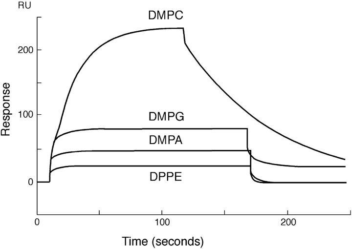
FIGURE 3.
Sensogram depicting the binding of PDC-109 to different lipid monolayers. PDC-109 was passed over different lipid monolayers (DMPC, DMPG, DMPA, and DPPE), containing 20% (wt/wt) cholesterol at 20°C. The concentrations of PDC-109 used were 0.05 μM, 20 μM, 100 μM, and 150 μM, respectively, for DMPC, DMPG, DMPA, and DPPE.

