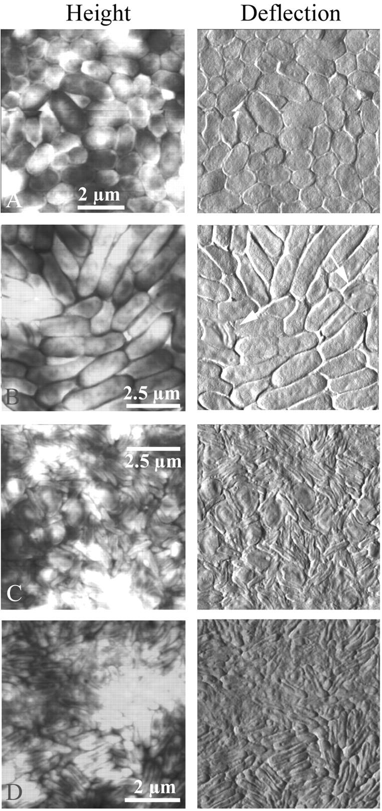
FIGURE 3.

The life cycle of Bdellovibrio in a population of E. coli was imaged by atomic force microscopy in contact mode (height and deflection images). Solutions of predator and prey were mixed and deposited on smooth, small-pore filters over DNB plates. At various times over several days, filters were removed to the AFM and imaged. (A) Prey E. coli grew quickly on the filters to form a continuous layer of cells. The total depth range of the height image was only ≈200 nm, compared to a cell height of ∼400 nm, which indicated that the cells were all roughly the same height and were therefore growing next to but not on top of each other. (B) A few invaded cells, or bdelloplasts, were initially present in the population of healthy cells. (C) Given a few days to hunt in the monolayer of prey, Bdellovibrio invaded every cell, and only bdelloplasts and predators were seen. (D) Eventually only the predator bacteria and some cell debris remained. The total depth range of the height image was ≈150 nm. The exact time elapsed between different stages depended upon the concentration and vigor of the two initial bacterial cultures, as well as the location on the filter that was imaged.
