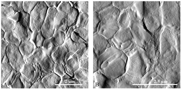
FIGURE 4.
High-resolution AFM images of a mixed E. coli-Bdellovibrio system, taken in contact mode (deflection). Several bdelloplasts were visibly mixed in with healthy prey cells. The bdelloplasts were round or irregular in shape and had a smooth surface texture, whereas the uninvaded cells generally had a more oblong shape and a rough surface texture. Furthermore, the growing Bdellovibrio were visible inside of many of the bdelloplasts as smaller, oblong, or coiled shapes. Arrows indicate bdellovibrios that were in the process of attacking prey cells.

