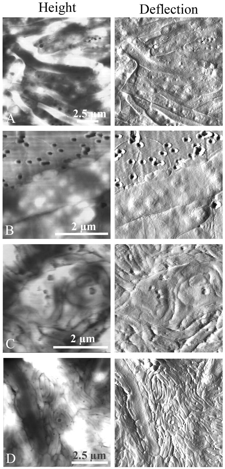
FIGURE 6.

The life cycle of Bdellovibrio in a population of Aquaspirillum serpens imaged by atomic force microscopy in contact mode (height and deflection). Again, solutions of predator and prey were mixed and deposited on smooth, small-pore filters over DNB plates, and at various times over several days, filters were removed to the AFM and imaged. (A) Healthy prey Aquaspirillum were very long and lumpy, and unlike E. coli many grew in multiple layers on top of the filter. The total depth range of this height image was ≈400 nm, and in other fields was >600 nm, which indicated that the cells grew on top of each other. The presence of “lumps” in the uninvaded cells made bdelloplasts difficult to identify. (B) Two Bdellovibrio invaded the same Aquaspirillum. (C) A long coil of growing Bdellovibrio was visible inside of the Aquaspirillum bdelloplast, as proposed earlier from electron microscope images. (D) As with E. coli mixed monolayers, given a few days to “graze” on the prey, Bdellovibrio invaded and consumed every cell. One remaining spirillum was being attacked and consumed at the left side of the figure. Note that because the Aquaspirillum prey cells grew three-dimensionally on top of the filter, the predator cells were also piled on top of each other and the cell debris. The total depth range of this height image was ≈1.2 μm, and in other fields was at least 800 nm.
