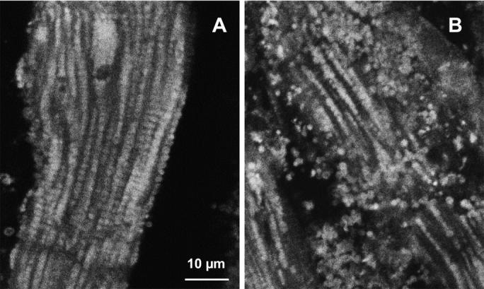
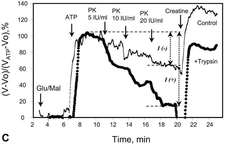
FIGURE 4.

Alterations of regular arrangement of mitochondria and of regulation of respiration in permeabilized cardiac fibers by trypsin treatment. (A and B) Confocal imaging of mitochondria using MitoTraker Green FM fluorescence. Permeabilized cells were preloaded with MitoTraker Green FM and fixed as described in Methods section. (A) Control fibers; and (B) after incubation with 1 μM of trypsin for 5 min at 4°C. (C) Representative oxygen consumption traces showing changes of metabolic channeling of endogenous ADP from Ca,MgATPases to mitochondria. For visualization of metabolic channeling, PK-PEP competitive enzyme method was used before and after treatment of permeabilized cardiac fibers with trypsin, leading to the disorganization of regular arrangement of mitochondria: the t-test. (The first derivative of oxygraph recordings of oxygen consumption are shown, directly showing the values of respiration rate.) ATP was added to the final concentration of 2 mM. (Thin line, control fibers. Thick line, fibers treated with 5 μM of trypsin for 15 min at 4°C.) I(−) and I(+) indicate inhibition of mitochondrial respiration by the competing PK-PEP system without (−) and with (+) trypsin treatment. At the end of each experiment, creatine (20 mM) was added.

