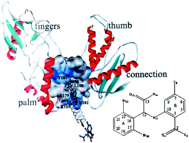
FIGURE 1.
A schematic representation of the 3D structure feature of p66 subunit of HIV-1 RT and the diagram of the SMD simulation system. The p66 subunit is shown in solid ribbon and α-APA in ball-and-stick model. The NNRTI binding pocket is represented with the VDW surface. During the SMD simulation, the bound α-APA is pulled away from the binding pocket using a harmonic potential symbolized by an artificial spring that is connected to the C12 atom of α-APA (labeled in green color). This pulling potential moves with a constant velocity of 0.02 Å/ps in the direction indicated by the arrow. Three snapshots of α-APA in the unbinding pathway are shown, representing the initial state, the state passing through the entrance of the binding pocket, and the final state in the SMD trajectory, respectively. This picture was generated using WebLab ViewerPro program (MSI, 1998) and rendered using the POV-Ray program (Pov-Ray-Team, 1999). The numbering of α-APA is shown in the schematic diagram of α-APA.

