
FIG. 3.
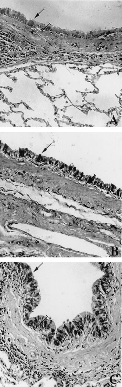
Goblet cells and mucus production at the bronchial epithelial layer of S. venezuelensis-infected rats. Sections of lung tissue from uninfected rats (A) and from rats 5 days after single infection (B) or 5 days after the last infection for multiple-infection rats (C). Tissue was fixed with buffered formalin and was embedded in paraffin, and 5-μm sections were stained with Alcian-Blue Safranin, pH 2.5. Note the increased number of goblet cells filled with mucus (dark areas indicated by arrows) on the epithelial layer and the epithelial layer (labeled with a bracket) after single and multiple infection with S. venezuelensis. Magnification, ×200.