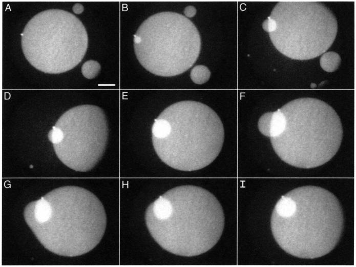
FIGURE 10.

Growth of a stacked disk. Temporal sequence of fluorescence micrographs show structures formed from monolayers of DPPC/dchol 60/40 (mol/mol) at 52 mN/m. Images were recorded at the following times after the initial image: (A) 0.0 s; (B) 15.0 s; (C) 68.8 s; (D) 73.7 s; (E) 87.8 s; (F) 133.0 s; (G) 133.1 s; (H) 133.2 s; and (I) 133.4 s. Scale bar, 50 μm.
