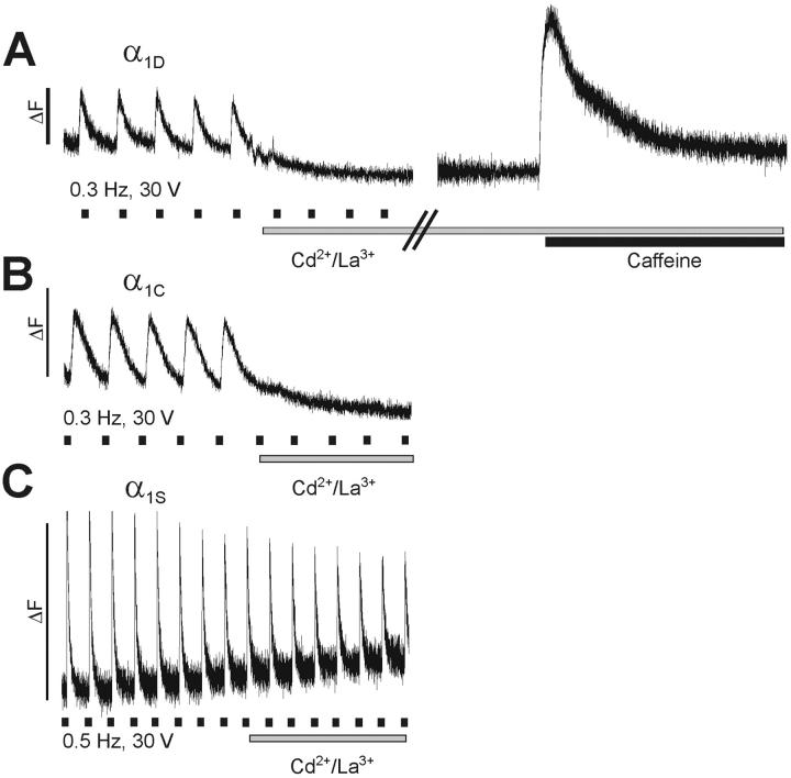
FIGURE 3.
Restoration of action potential-induced Ca2+ transients by GFP-α1D in dysgenic myotubes. Field stimulation at low frequency (30 V, 1 ms, 0.3 Hz/0.5 Hz) evoked rapid Ca2+ transients in myotubes expressing GFP-α1D, GFP-α1C, or GFP-α1S. Bath application of 0.5 mM Cd2+/0.1 mM La3+ immediately stopped the activation of transients in myotubes expressing GFP-α1D (A) and GFP-α1C (B), indicating that the transients were dependent on Ca2+ influx through the L-type channels. Addition of 6 mM caffeine (A, right trace) triggered a strong Ca2+ transient, proving that SR Ca2+ release was still functional during the Cd2+/La3+ block. (C) Persistent Ca2+ transients after addition of Cd2+/La3+ in myotubes expressing GFP-α1S characterizes skeletal-type EC-coupling, which is independent of Ca2+ influx. In contrast, GFP-α1D restored action potential-induced Ca2+ transients with cardiac characteristics. Marks underneath the traces indicate the approximate times of stimulation; bars indicate periods of Cd2+/La3+ and caffeine application.

