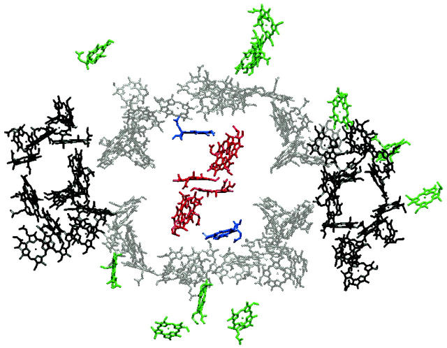
FIGURE 1.
Structure of a monomeric unit of PSI. Only the 96 chlorophyll molecules are drawn as Mg-chlorin rings, lacking the phytyl tail. The Chls in the RC are colored in red and the two linker Chls are in blue. The other antenna Chls are sectioned into three parts: central (gray) and peripheral (black) coordinated by protein units PsaA and PsaB (Jordan et al., 2001). The Chls in green are antenna Chls coordinated by other protein units. P700 was calculated to be a single Chl a located at the center of the RC in Article I.

