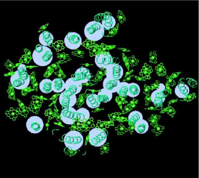
FIGURE 2.
Field model of the protein. Within this model, the protein medium (i.e., the medium with the refractive index of n = 1.2) is represented with a set of cylinders. The cross-section of these cylinders is shown with white circles. The real location of the trans-membrane part of α-helices in PSI is shown in turquoise, with the tube representation. Chlorophylls are presented in green as Mg-chlorin rings, lacking the phytyl tail. Chlorophyll Mg atoms are shown in van der Waals representation.

