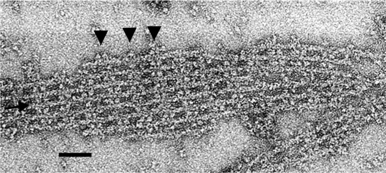
FIGURE 2.

Electron micrograph of a negatively stained actin-aldolase raft. This raft has an interstitial filament (see arrow), which is cross-bridged to its neighbors. To the right of the inserted filament, the two neighbors are cross-bridged to each other. The edges of outer filaments are decorated with aldolase in some places (see arrowheads). Scale bar = 500 Å.
