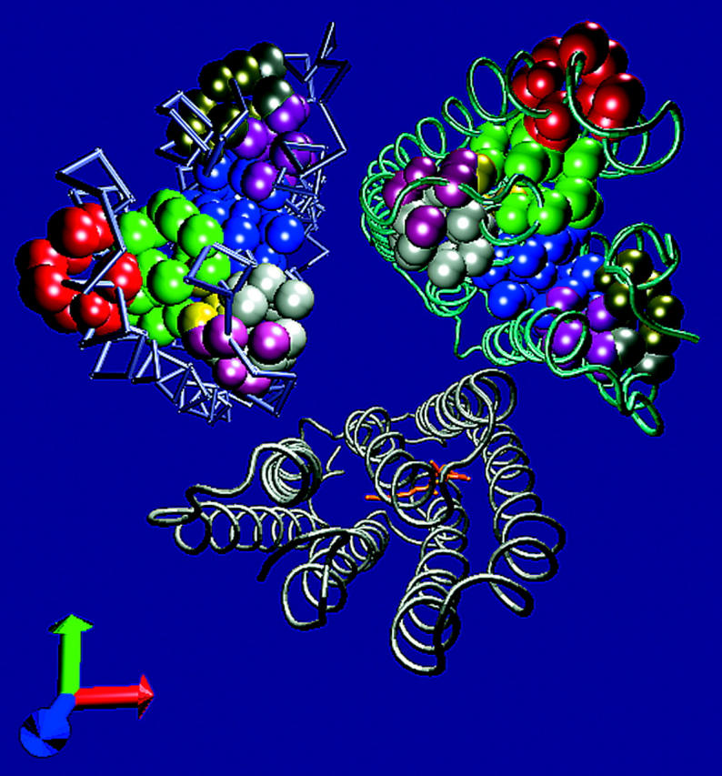
FIGURE 2.

The cavities on the cytoplasmic side of the BR. The atoms, which border the cavities of the cytoplasmic side of the BR, are presented as VDW spheres on the helical structure of the protein trimer structure 1BRR. The cavities, which were found on subunit C, are presented on subunits B and C, whereas only the retinal residue is presented on subunit A for orientation. See Table 3 for the properties of the cavities.
