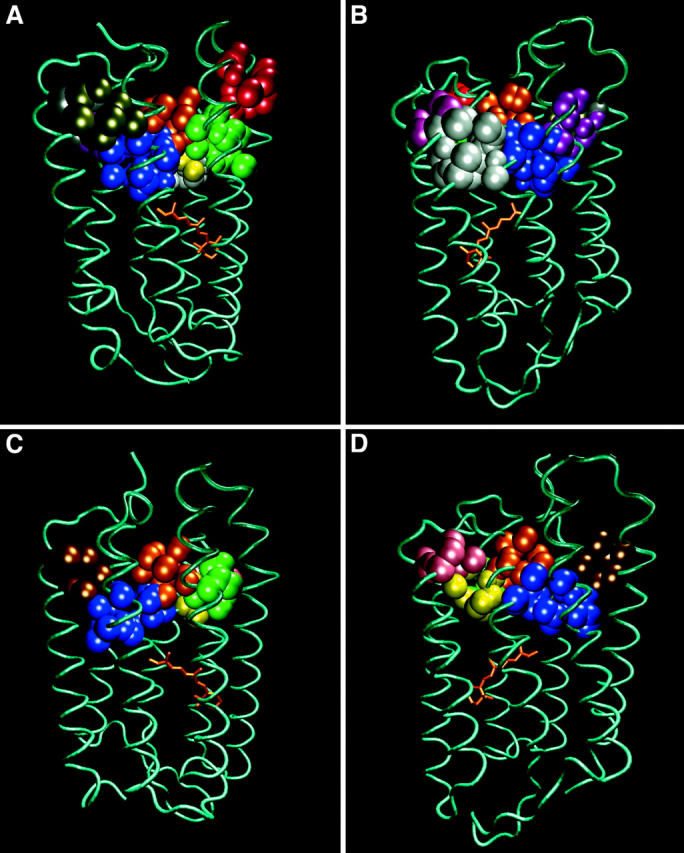
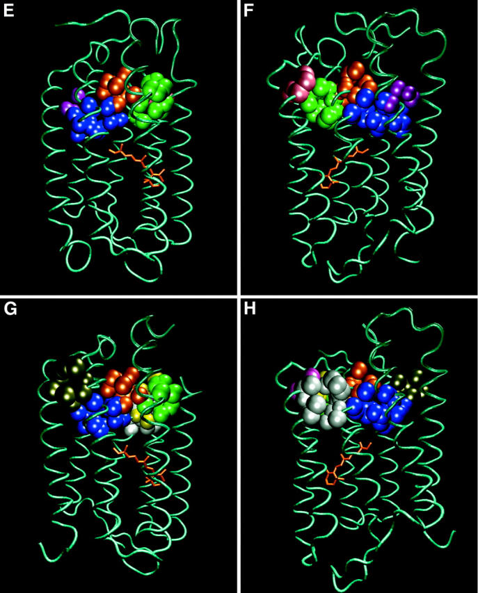
FIGURE 3.


A comparison of the shapes and sizes of the 10 cavities located in the cytoplasmic section of bacteriorhodopsin, as calculated for four ground state structures of the protein. Each pair of frames depicts the position of the cavities, colored as defined in Table 2. The orientation of the protein can be deduced by the location of the retinal molecule. Frames A and B: 1BRRC. Frames C and D: 1QHJ. Frames E and F: 1CWQA. Frames G and H: 1C3W.
