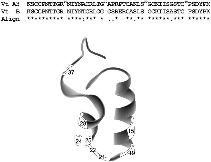
FIGURE 1.
Comparison of the amino acid sequence of viscotoxins A3 and B, indicating the differences in amino acid positions, as well as their alignment (Clustalw). The three- dimensional structure of viscotoxin A3 (PDB code 1ED0) is also represented, showing the location of the amino acid residues that are different between the two proteins.

