FIGURE 6.
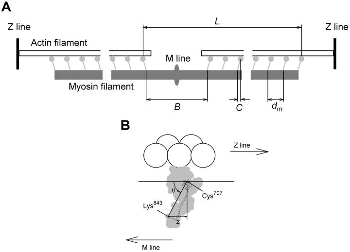
Sarcomeric location and conformation of myosin head domains. (A) Myosin heads (light gray) with head-rod junctions (Lys843) connected to the myosin filament backbone (dark gray) at axial periodicity dm, except for the central bare zone of length B. The center of mass of each level of myosin heads is displaced from its head-rod junction by a distance C. The interference distance (L) is defined in the text. (B) Myosin head (light gray) with catalytic domain (residues 1–707 of the chicken skeletal myosin heavy chain sequence) attached to an actin monomer (white sphere) in the conformation of Rayment et al. (1993a,b), and light chain domain (residues 707–843 of the myosin heavy chain (dark gray) and both light chains) rotated around Cys707 so that the Cys707–Lys843 axis makes an angle θ with the filament axis. z denotes the axial displacement of the catalytic domain (with Cys707 as reference residue) with respect to Lys843.

