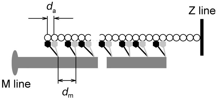
FIGURE 8.
Sarcomeric location of myosin heads; model with long-range order of the actin filament. The two heads of each myosin (black and light gray) bind to adjacent actin monomers (circles) with axial periodicity da, but share a head-rod junction with the axial periodicity (dm) of the myosin filament (dark gray). Only half the sarcomere is shown; axial intensity distributions were calculated from this model under the assumption that the sarcomere was symmetrical across the M-line.

