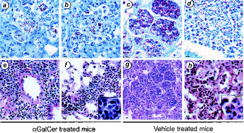
FIG. 5.
αGalCer treatment reduced pulmonary injury. BALB/c mice were infected with M. tuberculosis and treated with αGalCer (a, b, e, and f) or vehicle (c, d, g, and h). After 4 weeks, mice were sacrificed and the lungs were removed and fixed in formalin. Paraffin-embedded sections were stained with Fite-Faracco stain for AFB (a to d) or with hematoxylin and eosin (e to h). The CFU for this experiment are depicted in Fig. 4. Although both groups of mice had significant pulmonary infiltrates 4 weeks after inoculation, the αGalCer-treated group tended to have greater numbers of lymphocytes within the lesions (e, f, and panel f inset), whereas the vehicle-treated mice had more areas of necrosis and accompanying polymorphonuclear leukocyte infiltrates (g, h, and panel h inset). Bar, 10 μm.

