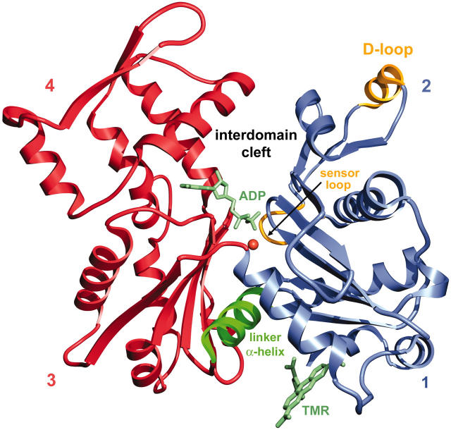
FIGURE 1.
Ribbon representation of the structure of TMR-actin in the ADP state. The two major domains on each side of the nucleotide cleft are shown in blue (subdomains 1 and 2) and red (subdomains 3 and 4). In F-actin subdomains 1 and 3 point toward the barbed end of the filament and subdomains 2 and 4 are directed toward the pointed end. Shown in yellow are the loop containing the methylated His-73 (sensor loop), which plays a key role in transmitting conformational changes from the nucleotide site to subdomain 2, and the D-loop, which changes conformation between ATP and ADP-actin. In green is the α-helix from Ile-136 to Gly-146, which serves as a hinge for rotation between the two major domains.

