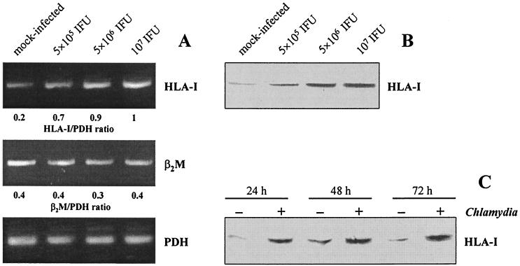
FIG. 1.
HLA class I expression in synovial fibroblasts infected with C. trachomatis D. (A) HLA-I and β2M mRNA levels in cells infected with various doses of chlamydiae. RT-PCR analysis was conducted on total RNA extracted at 24 h after infection. (B) Immunoblot analysis of HLA-I protein in cells infected with various doses of chlamydiae. Total cell lysates were prepared at 48 h after infection. (C) Time course of HLA-I production determined by immunoblotting. Fibroblasts were infected with 5 × 10⁶ IFU/well.

