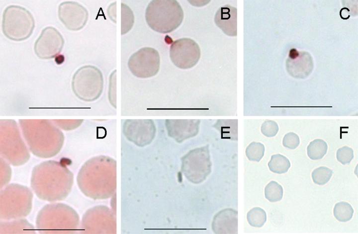
FIG. 6.
Expression of MSA-2 proteins by sporozoites at the time of attachment to erythrocytes. Smears of erythrocyte cultures initiated with sporozoites were incubated with specific mouse antiserum against MSA-2a (A), MSA-2b (B), MSA-2c (C), monoclonal antibody 23/10.36.18 against MSA-1 (D), or negative-control mouse antiserum against A. marginale MSP-2 OpAG3 (E). Erythrocyte cultures treated identically but with the use of extracts from uninfected larvae were incubated with specific mouse antiserum against each MSA-2 protein; anti-MSA-2a is shown in panel F (magnification, ×63,000). The reaction was visualized with AEC, which results in brown staining. Bars = 10 μm.

