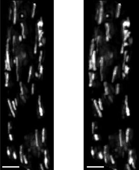
FIGURE 2.
Mitochondrial accumulation of TMRE. A high resolution stereo-pair fluorescence image of a toad stomach smooth muscle cell loaded with TMRE. Each image is a composite consisting of a projection of a three-dimensional image stack onto a single plane. The discrete localization of the dye to the mitochondria and the arrangement of the mitochondria in alignment with the long axis of the cell are apparent. Differences in mitochondrial FI, and therefore mitochondrial ΔΨm, are clear. The scale bars are 5 μm.

