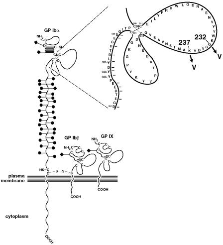
FIGURE 1.
The GPIb-IX complex. The drawing depicts the structural domains of the three subunits of the GPIb-IX complex along with an exploded view of the disulfide loop region of GPIbα. The mutations used in this study, K237V and Q232V, are highlighted. Solid circles and diamonds attached to the stalks represent O-linked and N-linked carbohydrates respectively.

