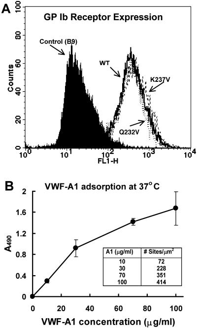
FIGURE 2.
(A) Cell surface expression of GPIbα (WT, Q232V, and K237V) on transfected CHO cells: The cells were incubated with FITC-conjugated mouse monoclonal antibody AK2 in 5% HSA solution for 60 min. The expression levels were detected using FACScan flow cytometry. CHO cells expressing GPIbβ-IX (without α-subunit) were used as control. (B) Adsorption of VWF-A1 on glass coverslips at 37°C. The absorbance values are averaged from two experiments performed in triplicate.

