FIGURE 4.
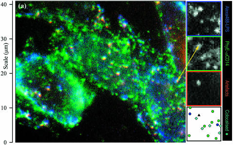
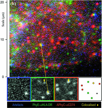
Color-merged images of (a) CHO-CD14 cells labeled with Alexa 488-LPS (blue) and PhyE-IgG(CD14) (green) and imaged with suitable filters and exposure times; the red channel detects artifacts that appear as white spots, and (b) M1DR1/Ii/DM cells labeled with PhyE-IgG(DA6.231) (green) and APhyC-IgG(BU45) (red); artifacts are detected using the blue channel. Colocalized spots and artifacts show imperfect registration due to chromatic aberration before correction. Insets are the separate images (shown by the edge colors) from a small area; the final inset is a map of the chromatically corrected fitted spot positions: an artifact (triple correlated) is shown as a black triangle, isolated spots appear as circles, correlated blue/green (Alexa488-LPS and PhyE-antiCD14) spots in a as cyan diamonds, and a correlated green/red (PhyE-antiHLA-DR and APhyC-antiCD74) spot in b, where it is marked by a short arrow, as a yellow diamond. Correlated spots are plotted at the coordinates of the green spot.


