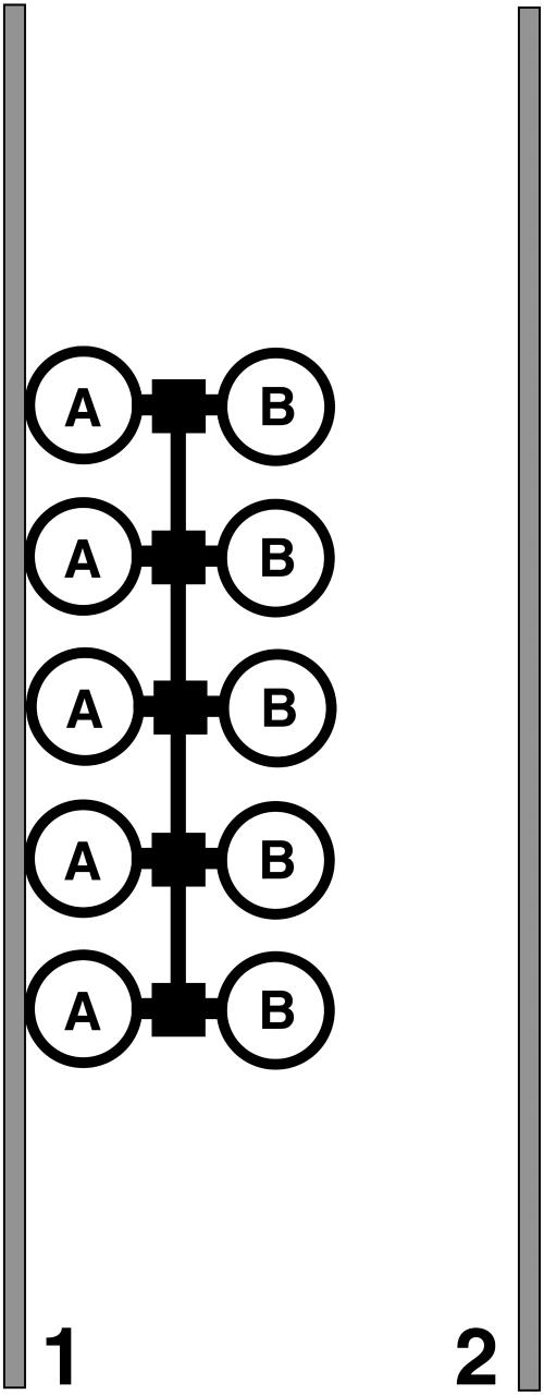
FIGURE 1.
Model for binding of H-NS to DNA. The two identical DNA binding domains (A and B) of each H-NS dimer (large circles) are directed in opposite directions and have the ability to interact independently with a stretch of double-stranded DNA (within the same or on another DNA molecule). Two stretches of dsDNA (1 and 2) are indicated in gray. H-NS dimers are held together through the oligomerization domain (small squares). In addition (a different region on) the same domain is responsible for oligomerization of adjacent H-NS dimers.

