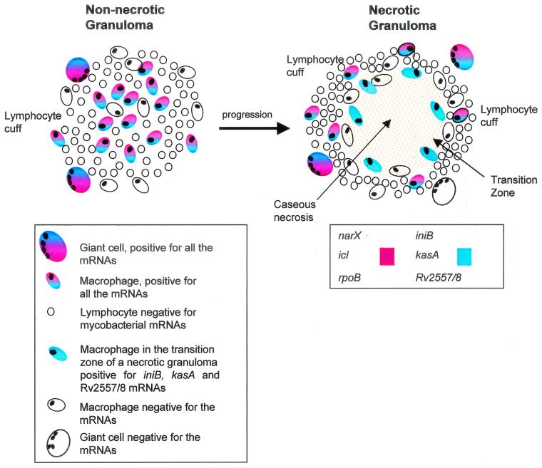
FIG. 4.
Schematic diagram of mycobacterial gene expression within a nonnecrotic tuberculous granuloma as it progresses and becomes necrotic. In a nonnecrotic granuloma consisting predominantly of macrophages, lymphocytes, and giant cells, mycobacteria expressing rpoB, narX, icl, Rv2557 and/or Rv2558, iniB, and kasA mRNAs were detected by using nonradioactive in situ hybridization. This nonnecrotic granuloma was also positive for M. tuberculosis DNA. As the granuloma progresses from a nonnecrotic to a necrotic state, the microenvironment changes, macrophages apoptose, and the bacteria slow their growth rate. The lymphocyte cuff of the necrotic granuloma contains a number of giant cells and isolated macrophages positive for the various mycobacterial genes. The transition zone of the necrotic granuloma still contains a small number of macrophages positive for Rv2557 and/or Rv2558, iniB, and kasA mRNA. However, the center of the necrosis is devoid of macrophages and mycobacterial gene expression but does contain M. tuberculosis DNA, as shown by DNA-DNA in situ hybridization.

