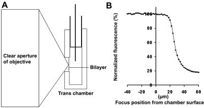
FIGURE 2.
Geometric considerations for optical imaging of planar lipid bilayers. The distance between the plane of the bilayer and the front surface of the trans chamber impacts optical measurements. The profile of the laser beam focused by a 70× high NA objective is cartooned in panel A. Due to its profile, the beam cannot be scanned very deeply into the hole without being scattered by the edges of the hole. This scattering causes a loss of signal intensity from a uniformly distributed fluorescent solution as the plane of focus is moved into the hole (panel B). To ensure that the entire surface of the bilayer is scanned (see Fig. 3), it is necessary to make the thickness of the trans-chamber wall 15 μm or less.

