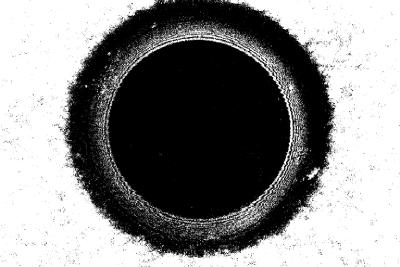
FIGURE 3.
Locating the bilayer. Bilayers are imaged using reflected 633-nm light to establish their location relative to the front surface of the trans chamber. A bilayer formed in a 100-μm diameter hole is shown in this image. The bilayer is the black inner circle and is ∼70 μm in diameter. The torus around the bilayer is shown by the white interference pattern and outer black region. The edge of the hole is the black to white transition. The depth of the bilayer in the hole is measured as the distance between the bilayer focal plane and the front surface of the trans chamber.

