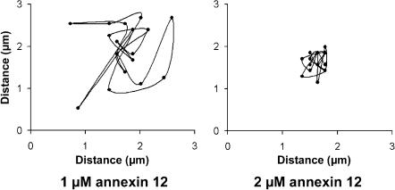
FIGURE 4.
A pixel-by-pixel analysis of RyR2 diffusion in the presence of 1 and 2 μM annexin 12. In the optical system used for these studies, pixels had a diameter of 0.12 μm and 20 data points were obtained at 3-s intervals. Thus, RyR2 were constrained to domains imaged by ∼±3.0 pixels over the course of 1 min in the presence of 2 μM annexin 12.

