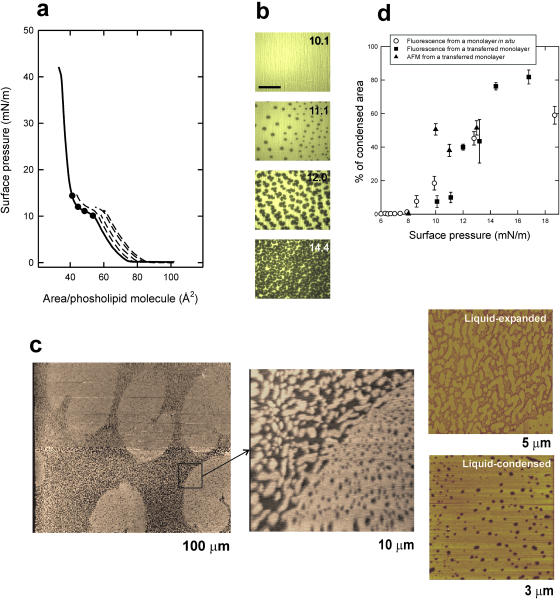
FIGURE 1.
(a) Surface pressure versus phospholipid molecular area of a DPPC monolayer spread and compressed on the surface of a pure water interface (continuous line) and isotherms (dashed lines) of films compressed to the pressures indicated by the black dots before being transferred at constant pressure onto glass coverslips, all done at 25 ± 1°C. (b) Epifluorescence images (scale bar, 100 μm) of DPPC monolayers containing NBD-PC (1% mol/mol) compressed to the indicated pressures before transfer as LB films onto glass coverslips, showing dark, probe-excluding condensed domains on a bright, probe-containing liquid-expanded background. (c) Contact mode SFM images from a DPPC monolayer containing NBD-PC (1% mol/mol) compressed to 11 mN/m and transferred to a freshly cleaved mica surface, at different magnifications (the width size covered by each micrograph is indicated). Contrast between condensed and expanded regions comes from differences in topology or film thickness as scanned by the SFM tip. (d) Quantitative analysis of the percentage of the films occupied by condensed domains at different surface pressures, calculated from DPPC monolayers containing 1% (mol/mol) NBD-PC and observed in situ by epifluorescence microscopy (open circles, data taken from Cruz et al., 2000) or upon transfer to LB supports and observation by epifluorescence (closed squares) or SFM (closed triangles).

