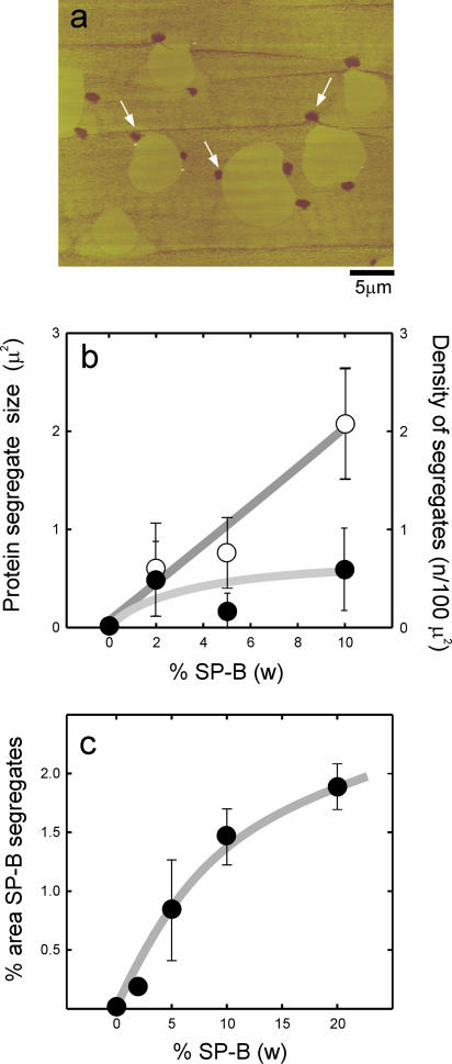
FIGURE 7.
(a) Contact mode SFM image of a DPPC film containing 5% (w/w) surfactant protein SP-B transferred onto mica from a monolayer compressed to 11 mN/m. White arrows indicate the presence of low patches, associated at the LE/LC borders, which are not present in supported films transferred from protein-free DPPC monolayers. (b) Quantitative analysis of the number (open circles) and size (closed circles) and (c) the total area of the patches versus the amount of protein in the films.

