FIGURE 9.
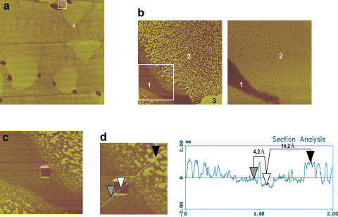
(a) Contact mode SFM micrograph showing topography of a 25 μm-width surface of a DPPC monolayer containing 5% (w/w) SP-B once compressed to 11 mN/m and transferred onto a mica support. (b) Magnification of a region centered at a single SP-B cluster, distinguishing the existence of three level surfaces (numbered 1–3) according to topography (left picture) and two regions (1–2) according to friction (right picture). (c) Material covering the protein cluster has been scratched in a square of 200 nm to uncover the mica surface. (d) Topographic profile through the different regions of the DPPC/SP-B film, showing estimated heights of protein cluster and lipid nanodomains with reference to the mica surface.

