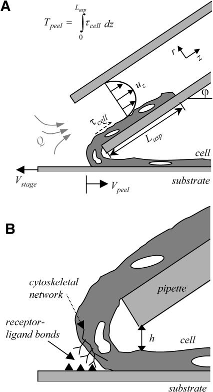
FIGURE 2.
Cell-peeling schematic in side view. A multinucleated muscle cell is aspirated into a large bore, cylindrical micropipette (of radius Rp). The shear stress, τcell, acting on the cell is integrated over the aspirated length of cell into the pipette, Lasp, to give the peeling tension Tpeel. At a flow rate of 10 μL/min, the Reynolds number for a 75-μm diameter pipette is 47.2. The motorized stage is translated to the left at Vstage in an effort to approximate or exceed Vpeel. A typical inclination angle of the pipette, ϕ, is 30–40°. (B) An enlarged view illustrates the height difference, h, established between the pipette and the top of the cell. Although h can vary between experiments, h is held constant once peeling begins, and is ∼5 μm. As sketched, receptor-ligand bonds between the cell and the substrate are broken during peeling. The amount of bending that occurs is influenced by ϕ, Tpeel, and the adhesion strength.

