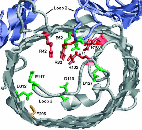
FIGURE 1.
Partial view of the x-ray structure of OmpF-trimer highlighting titratable residues in the constriction zone. Several loops have been truncated for clarity. Our calculations described in text predict residues colored in red to be positively charged at neutral pH and residues colored in green to be negatively charged at neutral pH. E296 colored in orange is expected to be protonated. Charge assignment of D127 was found to be sensitive to the value of dielectric constant assumed for the protein. Other assignments are not sensitive to assumption of dielectric constant. This figure was created with RasMol Version 2.6.

