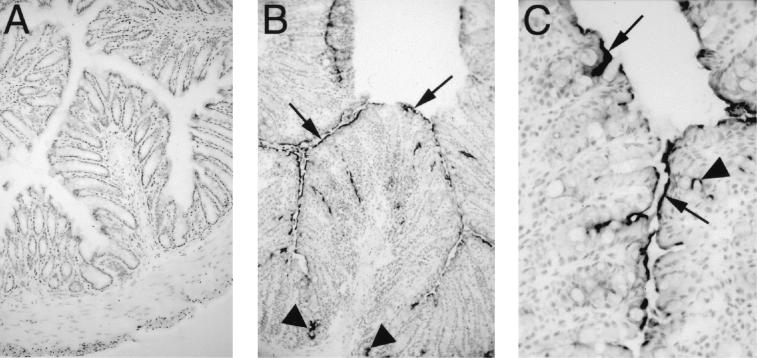
FIG. 2.
C. rodentium infection leads to iNOS expression by colonic epithelial cells. Immunohistochemistry for immunoreactive iNOS was performed on colonic tissues taken from uninfected mice (A), as well as mice at day 10 p.i. (B and C). Little if any immunoreactive iNOS was detectable in the colons of C57BL/6 mice prior to infection (panel A) (original magnification, ×100). By day 10 p.i., iNOS expression was seen along much of the mucosal surface (arrows) of infected C57BL/6 mice, including deep in the crypts (arrowheads) and by some scattered cells in the lamina propria (panel B) (original magnification, ×100). Under higher magnification, iNOS expression could be seen focused at the apical surface of many epithelial cells (arrows). A few lamina propria mononuclear cells were also found to express iNOS (arrowheads) (panel C) (original magnification, ×400).

