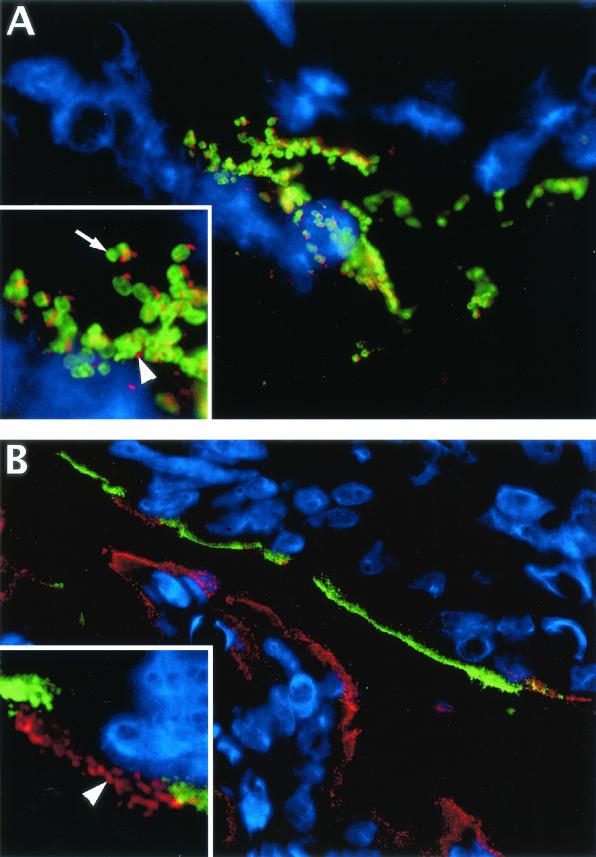
FIG. 7.
Immunofluorescence staining shows that infected cells in colonic tissue sections can be identified by their immunoreactivity for Tir but that Tir generally does not colocalize to cells expressing iNOS. (A) Colonic tissue section from a C. rodentium-infected mouse (day 10 p.i.) showing the lumenal surface of the colonic mucosa. Most host cells in this field of view (using DAPI to stain their nuclei blue) are superficial epithelial cells, while C. rodentium LPS is shown in green and Tir is shown in red. Large clusters of C. rodentium bacteria can be seen attached to these superficial epithelial cells during infection (original magnification, ×1,000). In the inset, at a higher magnification, one can see that Tir staining is not found inside the bacteria (arrow) but rather that it occurs outside of the bacteria (arrowhead), presumably translocated from the bacteria into the underlying epithelial cell and focused at the tip of actin pedestals. (B) This field of view shows the superficial colonic mucosa in both the upper and lower parts of the field, with the lumen in the center. By using the staining of Tir as a marker for infected cells, double immunostaining for Tir (red) and iNOS (green) shows that there is very little colocalization between cells expressing Tir and those expressing iNOS. The majority of the epithelial cells on the mucosal surface on the lower half of the picture are infected (Tir positive) but do not express iNOS, while the cells at the top express iNOS but are not infected and do not express Tir (original magnification, ×630). (Inset) At a higher magnification, one can see individual Tir foci in red (arrowhead) on the apical surface of one or two infected cells. These infected cells are the only iNOS-negative cells in a long stretch of uninfected epithelial cells expressing iNOS (green) at their apical surface.

