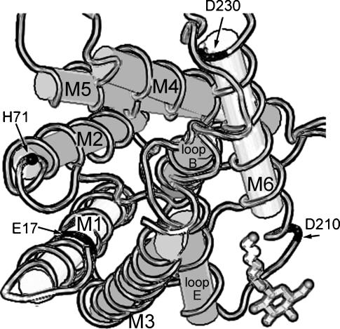
FIGURE 7.
Schematic diagram of a subunit of bovine Aquaporin-1, viewed from the cytoplasmic side. M1–M6 are membrane-spanning domains; loops B and E line the water pore within the individual subunit. The central cavity (putative ion pore) of the tetramer is lined by M2 and M5. Notations are Glu (E), Asp (D), and His (H). E17 in M2 and D210 and D230 near M6 (black fill) are homologous to the residues mutated in BIB (E71, D253, and E274). H71 (•) is postulated to interact with E17 in AQP1. The diagram is based on crystal structure data for bovine AQP1 (Sui et al., 2001) from the National Center for Biotechnology Information (NCBI) Molecular Modeling Database. File: MMDB 18789; PDB 1J4N; analyzed with Cn3D 3.0; available from http://www.ncbi.nlm.nih.gov. Structural similarities between BIB and AQP1 seem plausible but have not yet been demonstrated.

