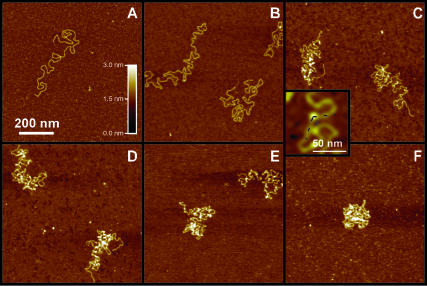
FIGURE 1.
AFM images of linearized pBR322 DNA molecules after exposure to increasing concentrations of Abf2p (ratio of Abf2p/bp in parentheses). (A) No Abf2p; (B) 1.5 μg/mL (1:20); (C) 3.5 μg/mL (1:8); (D) 7 μg/mL (1:4); (E) 15 μg/mL (1:2); and (F) 25 μg/mL (1:1). (Inset) Closeup image of a bend in the DNA backbone induced by the bound protein (bright dot).

