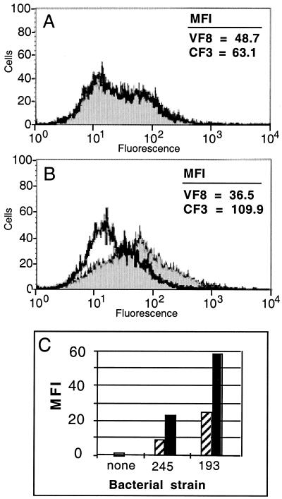
FIG. 1.
Measurement of cell surface CFTR expression on enterocytes obtained from ligated intestinal loops, before and after serovar Typhi infection. (A and B) CFTR-specific (CF3, filled histograms) or negative control (VF8, open histograms) antibodies were used for staining of live, intact epithelial cells followed by flow cytometric analysis. (A) Cell surface CFTR on uninfected enterocytes. (B) Cell surface CFTR on enterocytes after 1 h of infection with serovar Typhi. (C) Results of a similar experiment carried out with 15-min (hatched bars) and 1-h (solid bars) infection periods and using two other strains of serovar Typhi: H245 (245; identical to strain Ty2) and H193 (193). The values depicted represent the staining obtained with the CFTR-specific antibody CF3 corrected for background fluorescence obtained with the negative control antibody VF8. MFI, mean fluorescence intensity.

