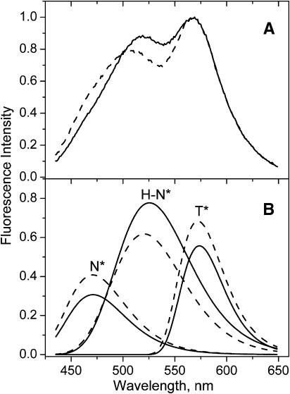
FIGURE 4.
Quenching of probe F emission in DOPC vesicles with TempoPC lipid. Fluorescence spectra (A) and corresponding deconvolution data (B) of probe F in DOPC vesicles in the absence (solid lines) and in the presence of 15% of TempoPC (dashed lines). Spectra are normalized at the T* band maximum. Experimental conditions are as in Fig. 3.

