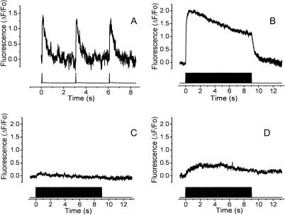
FIGURE 1.
Electrical field stimulation by single pulses elicits a rapid calcium signal in skeletal myotubes. The fluorescence was monitored by a photodiode attached to the side port of the microscope. The top trace shows the calcium transient and the bottom trace shows the electrical pulses. (A) Representative record of myotubes stimulated with 1-ms pulses at 0.33 Hz. (B) Control calcium transient induced by 400 pulses of 1 ms each at 45 Hz. The mean trace of 13 experiments is shown. (C) The same stimulation as in B but in the presence of 10 μM of TTX. The mean trace of 13 experiments is shown. (D) The same stimulation described in B; in cells preincubated with 30 μM of ryanodine. The mean trace of 10 experiments is shown.

